When you click on links to various merchants on this site and make a purchase, this can result in this site earning a commission. Affiliate programs and affiliations include, but are not limited to, the eBay Partner Network.
Thank you!
If you do not wish to have your item(s) delivered on data disc(s), I can provide them on a flash drive and other means as well. Just let me know if a disc does not work for you and we can discuss delivery by other methods.
COMBINING SHIPPING COSTS
Are you purchasing multiple items? I will: a) combine all invoices before payment and charge shipping equivalent to one item, or b) refund all shipping costs in excess of one item after payment.
All derivative (i.e. change in media; by compilation) work from this underlying U.S. Government public domain/public release data is COPYRIGHT © GOVPUBS.COM
$2.00 first class shipping in U.S. and rest of world.
Includes the Adobe Acrobat Reader for reading and printing publications.
Numerous illustrations and matrices.
Contains the following key public domain (not copyrighted) U.S. Government publication(s) on one CD-ROM in both Microsoft PowerPoint and Adobe Acrobat PDF file formats:
TITLE:
Selected Topics in Clinical Pathology, September 2005, 176 pages
SLIDE TOPICS, SUBTOPICS and CONTENTS:
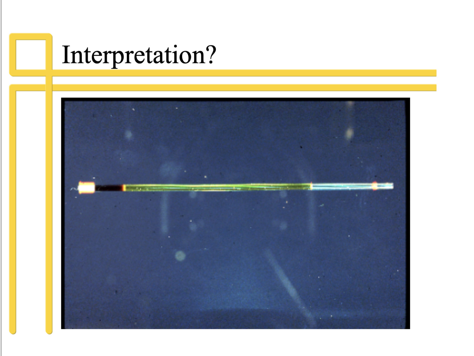
Selected Topics in Clinical Pathology US Army Laboratory Animal Medicine Residency Program Seminar 14-Sep-2005 The Blood Smear: What It Can Tell You Michael J. Topper, DVM, PhD Director, Clinical Pathology Safety Assessment Merck Research Laboratories Simple to Do! Basic Lab Equipment Microscope with a 100X oil lens Microhematocrit tubes and reader Refractometer Slides and coverslips Stains Immersion oil (Cell counter) Normal Microhematocrit Tube Interpretation? Examination of the Blood Smear Estimate white blood cell count Estimate platelet count Perform a differential white blood count Evaluate red and white cell morphology Observe etiologic agents Wedge-type Smears Staining the Blood Smear Wright’s stain Diff-Quik or other rapid stain New methylene blue stain Systematic Evaluation of the Smear Estimation of the White Blood Count With practice the WBC can be intuitively estimated as decreased, within reference range or increased; Or can be estimated by multiplying the average number of leukocytes per high dry field (40X) by 1500. Counting Chamber Counting Grid Platelet Examination Assume adequate numbers of platelets if an average of 8-29 per 100X oil field; Or estimate by counting the average number of platelets per 100X oil field and multiplying by 15,000. Erythrocyte Morphology Erythrocyte cell morphology is evaluated under oil at 100X in the same area of the smear in which the differential was done. Check for changes in size and shape, polychromasia, parasites, or inclusions. Erythrocyte Morphology Agglutination Anisocytosis Macrocytes Microcytes Polychromasia Hypochromia Erythrocyte Morphology Poikilocytes Spherocytes Basophilic stippling Heinz bodies Nucleated erythrocytes Parasites Classification of Anemia Normocytic, macrocytic or microcytic Normochromic or hypochromic Regenerative or nonregenerative Leukocyte Morphology Leukocyte cell morphology is evaluated under oil at 100X in the same area of the smear in which the differential was done. Look for signs of toxic change in neutrophils, or any unusual features of cell size or shape, parasites, or inclusions. Leukocyte Morphology Toxic change Neutrophil hypersegmentation Neutrophil hyposegmentation Asynchronous maturation Immunocytes Immature leukocytes Toxic Changes in Neutrophils Cytoplasmic basophilia Toxic granulation Foamy cytoplasm Doehle bodies Giant neutrophils, bands and metamyelocytes Case 1 - Dog History and Physical Exam: 2 year old mixed breed presented with lethargy, weakness, icterus and pale mucous membranes Laboratory Data: Hematocrit: 15% Plasma color: Very yellow Plasma protein: Normal Case 1 - Dog Blood Smear Evaluation: Leukocytes WBC: 50,800 Differential: Mature neutrophilia and monocytosis Tilted Drop of Blood Case 1 - Dog Blood Smear Evaluation: Erythrocytes Polychromasia Anisocytosis Spherocytosis Autoagglutination Nucleated red blood cells Case 1 - Dog Diagnosis: Autoimmune hemolytic anemia Case 2 - Horse History and Physical Exam: 12 year old Appaloosa gelding presented with depression, dehydration and icterus. Laboratory Data: Hematocrit: 25% Plasma protein: 8.0 g/dl Color: Plasma-orange & blood-chocolate Case 2 - Horse Blood Smear Evaluation: Leukocytes WBC: 11,900 Differential: Mature neutrophilia and lymphopenia Case 2 - Horse Blood Smear Evaluation: Erythrocytes Heinz bodies Eccentrocytes Case 2 - Horse Diagnosis: Acute intravascular hemolytic anemia due to ingestion of red maple leaves Case 3 - Dog History and Physical Exam: 5 year old female pit bull terrier presented with vomiting, fever, distended abdomen, dehydration. Laboratory Data: Hematocrit: 31% Plasma protein: 8.5 g/dl Case 3 - Dog Blood Smear Evaluation: Leukocytes WBC: 150,000 Differential: Neutrophilia with severe left shift and monocytosis Morphology: Toxic changes Case 3 - Dog Blood Smear Evaluation: Erythrocytes Normocytic Normochromic Case 3 - Dog Diagnosis: Inflammatory leukogram with anemia of chronic disorders due to a pyometra Case 4 - Dog History and physical exam: 6 year old German shepherd dog presented with lethargy, pale mucous membranes Laboratory Data: Hematocrit: 20% Plasma protein: 7.5 g/dl Case 4 - Dog Blood Smear Evaluation: Leukocytes WBC: 2,336 Differential: Neutropenia, eosinopenia, lymphopenia Morphology: Morula of Ehrlichia canis in monocytes Laboratory Evaluation of Hepatobiliary and Renal Disease Michael J. Topper, DVM, PhD Director, Clinical Pathology Safety Assessment Merck Research Laboratories Chemistry Patterns Tests can be grouped together on the basis of body system or physiologic process Grouping tests into common parameters is the best way to interpret chemistry data as it enables pattern recognition Patterns of change within and among these groups can provide useful diagnostic information about disease involvement of various organ systems Chemistry Tests Groupings Electrolytes: Na, K, Cl Acid/base: Bicarb, AG Minerals: Ca, Phos, Mg Protein: TP, Albumin Globulin, A/G ratio. Kidney: BUN, Creat Pancreatic: Amylase, Lipase, TPLI Lipid: Chol, Triglycer Iron: Iron, TIBC, % saturation of transferrin Liver Parameters: Leakage Enzymes - ALT, AST, SDH Cholestasis - AP, GGT Liver Function - Total & Direct bili, BA, NH3 CHO: Glucose, Fructosamine, Gly. Hb Muscle: CK, AST, ALT, LDH Reference Ranges Reference ranges vary considerably from one laboratory to another, and are dependent on the methodology, reagents and instrumentation utilized Published "normal" values may not be valid for results generated by your lab Do not compare one laboratory's results to another laboratory's reference intervals Reference Ranges Small deviations outside the reference interval may not be significant Clinically healthy animals may have mildly reduced or increased analyte values compared to a reference interval, but the values are actually normal for that animal Depends on the analyte, e.g. mild elevations in liver enzymes may not significant, however, electrolytes are maintained within fairly narrow limits and elevations in these are more likely to be clinically significant Hepatocellular Leakage Enzymes Useful in detecting injury to liver parenchymal cells with increased serum activity representing enzyme leakage through damaged cell membranes AST: Used in small and large animals. Liver as well as skeletal muscle and erythrocytes ALT: Used in small animals only. Largely liver-specific, but can also increase in severe myopathies and hemolysis SDH: Liver specific in nearly all species. Used in large animals in place of ALT Disease Effects on Serum Levels Serum levels depend on both the number of cells affected and the severity of injury to individual cells to allow “leakage” Serum levels do not correlate with reversibility as diffuse hypoxia (reversible injury) may result in greater serum activity than end-stage cirrhosis (irreversible injury) Increases are not specific with regard to the nature of the injury Increased Serum Levels "Primary" hepatic disorders Inflammation: Viral, bacterial, fungal, immune-mediated, idiopathic Intoxications: Drugs, chemicals, plants Neoplasms: Hepatocellular and bile duct carcinomas, metastatic neoplasia Disorders with secondary hepatic effects Circulatory: Heart failure, shock, severe anemia, portosystemic shunts, septicemia Metabolic: Diabetes mellitus, Cushing's disease, and idiopathic lipidosis, acute pancreatitis Alanine Aminotransferase - ALT Located in the cytoplasm Useful as a specific indicator of hepatocellular injury in most small animals, but not Guinea pigs Half-life is 2-5 days in dogs and < 24 hours in cats Following acute hepatic injury, activity peaks at about 48 hours and then begins to decrease Increases in the enzyme occur due to cell damage (increased membrane permeability or necrosis) and induction (increased synthesis) ALT ALT is virtually liver specific in dogs, cats, rabbits, rats and primates Some increases are possible in severe muscle diseases of the dog and cat due to release of enzyme from this tissue; may see increase in mouse if squeeze when pick up ALT is found in the liver, muscle (cardiac and skeletal), kidneys, and erythrocytes Increased ALT Artifact: Hemolysis in the cat Drugs: Anticonvulsants increase ALT 4 x normal Corticosteroids increase ALT 2-3 x normal Any drugs that can cause hepatotoxicity can result in increased ALT levels, e.g. tetracycline in cats, caparsolate in dogs, acetaminophen. (Certain drugs may decrease ALT (and AST) activity, by impairing activation of vitamin B6 to P5P, e.g. cephalosporin, cyclosporin, isoniazide) Increased ALT (Liver Disease) Both primary and secondary hepatic disease can cause increased ALT levels, if altered cell membrane permeability or necrosis occur Usually ALT values exceed AST values Bile duct obstruction will increase ALT due to the toxic effects of retained bile salts on hepatocytes Trauma will often increase ALT levels, even without morphologic evidence of cell injury. Increased ALT (Muscle Disease) In small animals with severe muscle injury (ischemic myopathy in cats, muscular dystrophy in dogs), ALT will increase with CK and AST The increases in ALT are usually less than increases in AST in primary muscle disease and SDH values will be normal Aspartate Aminotransferase - AST AST is not organ specific. It is found in liver, cardiac and skeletal muscle, erythrocytes, renal epithelial cells and brain tissue Located in the cytoplasm and mitochondria as different isoenzymes The half life is 5-12 hours in the dog, 77 minutes in the cat, and 1-2 days in large animals More sensitive than ALT in detection of hepatobiliary disease in dog/cat, but less specific Increased AST Artifact: Hemolysis or leakage from cells can cause erroneously high values Drug effects: Anticonvulsants and corticosteroids may induce synthesis in dogs Physiologic effects: In horses, exercise can increase serum activity as much as 30% Increased AST (Disease) Myopathies: Muscle trauma/necrosis, malignant hyperthermia and muscular dystrophy may result in marked increases Liver disease: AST will increase in liver disease with the same causes as for ALT. Increased levels seen with hepatocellular injury often aren't as high as those seen with muscle damage. CK levels are normal unless there is concomitant muscle disease. Other liver specific enzymes (SDH) would also be increased Sorbitol Dehydrogenase - SDH Highest concentration in the liver. Cytoplasmic enzyme with a short half life (12-24 hours), but is not a stable enzyme Very specific indicator of liver disease in all species, although increases can occur with primary or secondary liver disease Increases occur within 24 hours of liver injury SDH is the enzyme of choice for detecting hepatocellular injury in horse and cattle Other “leakage” enzymes Lactate dehydrogenase (LDH) Wide tissue distribution, primarily skeletal and cardiac muscle and liver Liver isoenzyme is major component of total LDH in the dog, but little diagnostic value Arginase Liver specific in dog and cat, located in mitochondria, short half-life (<12 hours) Not routinely evaluated Cholestasis Parameters Serum alkaline phosphatase (AP) and gamma glutamyl transpeptidase (GGT) serve as indicators of cholestasis (impairment of bile flow) When severe enough, cholestasis will result in elevations of bilirubin, however, AP and GGT are more sensitive for cholestasis AP is less specific for cholestasis than GGT Alkaline Phosphatase- AP Sensitive indicator of cholestasis in the dog (increases before bilirubin), however it is non-specific as corticosteroids (exogenous or endogenous "stress") induce increases In the cat, it is a very specific indicator of liver disease, whereas in large animals, the enzyme is not very useful as it is non-specific In the rat, the intestinal isoenzyme is the predominant form and complicates interpretation as it increases with feeding and decreases with food deprivation AP Isoenzymes Four major isoenzymes of AP: intestinal (IAP), corticosteroid (CAP), bone (BAP) and liver (LAP) (also renal and placental) The corticosteroid isoenzyme is unique to dogs and is induced by corticosteroids (endogenous or exogenous) It takes approximately 10 days for CAP to be induced; therefore, initial increases in total AP with corticosteroid administration is due to increases in the LAP, not the CAP AP Isoenzymes Half-life varies with species & isoenzyme: LAP isoenzyme: dog - 66 hours, cat - 6 hours CAP isoenzyme: dog - 70 hours BAP isoenzyme: ? (but about 1/3 total AP) Placental, renal, and intestinal isoenzymes: dog - less than 6 min, cat - less than 2 min (do not really contribute to serum AP, except for intestinal isoenzyme in the rat) AP Isoenzymes Routine measurement of AP gives total serum activity without specificity as to source Isoenzyme measurement is commonly applied to canine samples with high AP to distinguish LAP and CAP Levamisole inhibits LAP and BAP Wheat germ lectin precipitates BAP and CAP Increased AP Drug effects: Glucocorticoids: In dogs, increased total SAP is due mainly to synthesis of the CAP isoenzyme. Marked increases are possible (50-100 fold). Total AP may remain high for three to six weeks, depending on the drug preparation administered (ie, short-acting vs. depot forms). Anticonvulsants: phenobarbital, primidone, phenytoin - mild to marked increases in total activity occur, due mainly to raised LAP isoenzyme Increased AP Age effect: AP activity in young, growing animals of all species may be 2 - 10 times higher than in adults, due to increased BAP isoenzyme Values decrease within 3 months of age and are within adult ranges by 15 months of age Increased osteoblastic activity (BAP isoenzyme): Primary and secondary hyperparathyroidism (2-3x inc) Osseous neoplasia (variable increase). Hyperthyroidism in cats: Mostly due to increased BAP with a lesser increase in LAP Increased AP Hepatobiliary disease - Increases occur in LAP In cats, AP increases in hepatobiliary disease, but the increase is less reliable and of lesser magnitude compared to the situation in dogs (feline hepatic tissue contains much less AP and serum half life is only six hours), therefore, any increases in AP in the cat are significant The wide range of SAP activities in normal cattle and sheep limits utility of SAP in these species Increased AP Extrahepatic cholestasis (bile duct obstruction): This causes very dramatic increases in AP and may occur before development of icterus, especially in the dog Intrahepatic cholestasis: Lesions that are primarily centrilobular generally cause only mild increases in AP while lesions affecting the periphery (periportal areas) of the lobule usually result in more dramatic elevations as a result of impaired bile flow Increased AP Neoplasia: Primary and metastatic liver cancer may increase AP due to localized cholestasis (may be only increase on chemistry panel) Acute hepatocellular injury: Mild to moderate elevations in AP are possible due to intrahepatic cholestasis associated with hepatocellular swelling (expect inc. in leakage enzymes) Hyperadrenocorticism - Levels vary from moderate to marked (up to 100- fold) Endogenous stress may increase AP 2-3 x normal Gamma Glutamyl Transferase - GGT Found primarily in microsomal locations in many tissues, but serum GGT primarily from the liver Liver: hepatocyte canalicular membrane Renal: brush border of prox tubules Pancreas: apical surface of acinar cells Used mainly as a sensitive indicator of cholestasis with a half life of 96 hours, except in rodents, as they have extremely low normal activity; 5’-nucleotidase is recommended as rodent cholestasis marker Increased GGT Drug effects: Increases in GGT occur secondary to therapeutic drugs causing cholestasis. Increases may also be seen with anticonvulsant and corticosteroid therapy Physiologic effects: Colostrum in all species, except for horses, contains high GGT concentrations. Increases in GGT occur within 24 hours of suckling and are a sensitive indicator of passive transfer Increased GGT GGT is a sensitive indicator of cholestasis in dogs and cats (but not in rodents). GGT increases may precede increases in AP In large animals, GGT appears to be an indicator of both hepatocellular injury, and cholestasis GGT is also a sensitive and specific indicator of biliary hyperplasia in large animals Clinical Tests for Liver Function Bile synthesis and secretion: bilirubin and bile acids Ammonia and amino acid metabolism: ammonia and BUN Protein synthesis: albumin, clotting factors, alpha & beta globulins Glucose homeostasis: glucose Lipid metabolism: cholesterol, triglyceride Bilirubin Assay Circulating bilirubin exists as conjugated (direct) and unconjugated (indirect) forms In the diazo reaction, the 1st step measures direct bilirubin, and 2nd measures total bilirubin, with unconjugated (indirect) being the difference No significance should be placed on the direct bilirubin when the total bilirubin is within the normal range Clinical icterus is observed when total bilirubin values exceed 1.5 mg/dL Delta Bilirubin Is conjugated bilirubin bound to protein Increases in serum when hepatic excretion of conjugated bilirubin is impaired (cholestasis) and the liver retains intact conjugation mechanisms It has a long half-life and is not excreted in the urine (as it is protein bound) Delta bilirubin may be responsible for a persistent bilirubinemia without bilirubinuria seen in some animals with cholestasis Increased Bilirubin Artifact: Hemolysis and lipemia (even mild) in the test sample will cause high bilirubin values Hemolysis: Overproduction of bilirubin saturates the liver’s capacity for uptake and conjugation, so unconjugated bilirubin is retained in the blood. With time, secretory function of liver becomes saturated and conjugated bilirubin “regurgitates” into the blood Increased Bilirubin Hepatic disease may cause increases in both unconjugated and conjugated bilirubin Increases in bilirubin in dogs often occurs after elevation of cholestatic enzymes (GGT, AP) due to the low renal threshold for bilirubin In large animals, increases in bilirubin are usually due to unconjugated bilirubin and bilirubin is only increased in cattle with very severe liver disease Increased Bilirubin Intrahepatic cholestasis due to either hepatocyte swelling or impaired secretion will result in bilirubinemia from higher conjugated than unconjugated bilirubin. In horses, a conjugated bilirubin >25% total bilirubin supports a diagnosis of cholestasis (as does bilirubinuria) Hepatic uptake and secretion may also be altered in sepsis and get hyperbilirubinemia Increased Bilirubin In horses, fasting will produce a hyper-bilirubinemia due to unconjugated bilirubin in the absence of significant liver disease Increases in bilirubin are noticeable within 12 hours of fasting and may reach levels as high as 10-12 mg/dL within 2-4 days of anorexia, with clinical icterus A similar mild increase in bilirubin (mostly unconjugated) can occur in anorectic cattle Increased Bilirubin Young animals, especially foals, often have jaundice, due to multifactorial causes, including hemolysis of fetal red blood cells, decreased liver uptake of bilirubin, immaturity of hepatic conjugation mechanisms and poor albumin binding Increased Bilirubin Inherited defects in hepatic uptake, conjugation and excretion of bilirubin occur in monkeys, sheep, and rats Southdown sheep have a defect (Gilbert’s) in bilirubin uptake, resulting in a fasting hyperbilirubinemia due to unconjugated bilirubin Corriedale sheep have Dubin-Johnson syndrome, a fasting hyperbilirubinemia due to conjugated bilirubin from defective excretion of conjugated bilirubin Bile Acids - BA BA are synthesized exclusively in the liver from cholesterol, and then are hydroxylated and conjugated to facilitate their role in fat digestion and absorption Conjugated BA are excreted into bile, stored in the gall bladder until ingestion of a meal stimulates their release Undergo efficient enterohepatic circulation that operates at 95% efficacy Determination of Bile Acids During fasting, BA are low and increase after a meal After a 12-hour fast, BA concentration is determined and again 2-hours after a small meal This exposes the portal vein to a large bolus of BA and tests its uptake capability Bile Acids Increased Hepatic disease Reduced functional mass (reduced uptake) Cholestasis (regurgitation back into blood) Portal shunts (decreased hepatic uptake) Decreased Ileal disease (dec absorption, inc fecal loss) Decreased synthesis (rare because of reutil) BA Limitations Many factors influence the enterohepatic circulation in normal animals BA can not discriminate one hepatobiliary disease from another, but some patterns are: Posthepatic obstruction: Very high fasting, with little change after a meal Shunts: Normal/slight inc fasting, but dramatic increase after a meal Little correlation between severity of histologic disease or degree of shunt and extent of BA inc Ammonia – NH3 Ammonia is a byproduct of GI protein catabolism and amino acid metabolism Readily diffuses thru the intestinal mucosa and enters portal circulation After hepatocyte uptake, it is detoxified by the urea cycle to urea or consumed in the synthesis of glutamine Increased Ammonia Hepatic synthetic failure (must be fairly advanced as urea cycle operates at only 60% capacity) Shunting of portal blood (sensitive since directly deposited into systemic circirculation) Deficiency of urea cycle enzymes or substrates Ammonia Limitations Critical sample collection and handling is necessary for accurate results Sample must be drawn into cold-heparinized tubes, immediately put on ice, and either assayed within an hour or quick-frozen (-20C) and assayed within 48 hours Diagnostic value improved with ammonia tolerance test Kidney Parameters Urea Nitrogen Creatinine Urine specific gravity (Proteins, minerals, electrolytes, acid-base balance, hematopoiesis) Glomerular Filtration Rate BUN and creatinine are indicators Decreases in GFR: Decreased renal perfusion (prerenal) Hypovolemia Cardiac dysfunction Loss of functional nephrons (renal) Combination of the above Other Tests of Glomerular Function Creatinine clearance for GFR Endogenous and exogenous Radioisotope and inulin clearance GFR, effective renal plasma flow (ERPF), filtration fraction (FF) Urinary protein 24-hr protein excretion; urine protein/urine creatinine ratio Tests for Tubular Function Urine specific gravity and osmolality Total urine solute concentration Water deprivation test Fractional clearance of electrolytes Expresses the proportion of a substance that is excreted in urine compared with that filtered through glomerulus Na is usually compared to creatinine Azotemia Defined as an increase in BUN and creatinine and can be due to prerenal, renal or post-renal causes Differentiation requires UA, physical examination and other diagnostic tests Uremia is term for clinical syndrome of renal failure with azotemia and other sequelae of inadequate renal function Prerenal Azotemia Due to a decreased GFR from circulatory disturbances causing decreased renal perfusion Dx with clinical signs (dehydration or hypovolemia), urinalysis (conc. urine and no evidence of renal tubule dysfunction) and response to therapy (fluid administration) Renal Azotemia Results from a decreased GFR when more than ¾ of nephrons are non-functional Due to primary intrinsic renal disease (GN, ethylene glycol) or secondary to renal ischemia from prerenal causes or from kidney damage from obstruction Renal Azotemia Isosthenuric urine UA evidence of renal tubular dysfunction High anion gap metabolic acidosis Inc Mg and K with oliguric and anuric renal failure Dog/cat: Dec K in cats with polyuric renal failure, Ca variable Post-renal Azotemia Secondary to obstruction or rupture of urinary outflow tracts Dx with clinical signs and ancillary diagnostic tests (radiographs, inability to catherize) Marked increases in K and Mg Can result in renal tubular dysfunction from impaired renal flow Urea Concentration Measured to screen for decreased GFR Urea nitrogen (BUN/SUN) Rats and dogs tight ranges while mice and monkeys have broad ranges Synthesized by hepatocytes from ammonia generated by protein degradation Urea is excreted by the kidneys, intestine (high in horses), saliva and sweat In ruminants, urea is excreted into the GI system where it is converted to amino acids and ammonia which are then used for protein production BUN BUN concentration is dependent on: Hepatic urea production Dec in liver disease (shunts) Inc with inc protein catabolism/digestion Renal tubular flow rate Freely filtered thru glomerulus and passively diffuses out of the tubules at a rate dependent on flow rate thru the tubules Remainder is excreted in the urine BUN Increased BUN Increased protein catabolism Increase protein digestion (esp. rodents) Decreased GFR Increased BUN with normal creatinine Increased prod. of urea (protein catabolism) Early prerenal azotemia Artifactual depression of creatinine (icterus) BUN Decreased BUN Decreased protein intake/anabolism Increased excretion due to polyuria Decreased production due to liver disease Creatinine Measured as a screen for GFR Produced as a result of normal muscle metabolism with phosphocreatine forming creatine then creatinine Minor from gut absorption after meat meal Freely filtered thru glomerulus and NOT reabsorbed in the tubules More reliable for GFR as it is not influenced by diet or protein catabolism Creatinine Increased creatinine: Artifact-in Jaffe technique both creatinine and non-creatinine chromogens (such as ketones) react with reagent Physiologic-higher in foals and heavily muscled horses Decreased GFR-best indicator in horses/rum. Increased production-minimal increase after a recent meat meal Creatinine Decreased creatinine: Artifact-with Jaffe reaction, severe icterus does this (lab needs reaction blank) Decreased production-loss of muscle mass or severe liver disease causes decrease creatine production Increased GFR-seen with portosystemic shunts and during pregnancy (inc cardiac output) Routine Urinalysis Physical properties: appearance, specific gravity Chemical properties: pH, protein, glucose, ketones, occult blood, bilirubin, leukocyte esterase reaction Sediment examination: RBC, WBC, epithelial cells, casts, crystals, organisms, miscellaneous Not very practical with rodents Sample Handling Unless the UA can be performed within a ½ hour of collection, refrigerate the sample Can be refrigerated for up to 12 hours, but warm to room temp before analyzing Warming allows some of the crystal precipitates to redissolve, and some of the chemical enzymatic reactions are inhibited by low temperatures and will yield false low or negative results Urine Color Normal urine is yellow due to presence of urochrome pigment Darkens with concentration and lightens with dilution Red/red brown: RBC, Hb or myoglobin Yellow-brown/green: bilirubin Cloudy: increased cells, crystals, mucus Specific Gravity Reflects the total solute concentration of urine (measurement of the density of urine compared to pure water) Used to assess the ability of the renal tubules to concentrate or dilute the glomerular filtrate Amount of any substance in urine must be interpreted in light of sp gr (e.g. 4+ protein more severe in 1.010 than 1.045 urine) Determine before Rx as fluids, diuretics, or glucocorticoids may alter Specific Gravity Urine pH Varies with diet and acid-base balance pathologic abnormalities of systemic acid/base balance pathologic abnormalities of tubular function with failure to excrete an acid load or failure to absorb bicarbonate dietary factors due to differences in dietary "acid load“ herbivores usually have alkaline urine carnivores tend to have acidic urine Age of specimen (CO2 loss into air raises the pH) Presence of contaminant or pathogenic bacteria (some convert urea to ammonia, raising pH) Urine pH Knowledge of the urine pH is important in interpreting urine sediment findings Erythrocytes, leukocytes, and casts tend to disintegrate in alkaline urine (pH > 8.0) In addition, precipitation of urine crystals in supersaturated urine is highly dependent on urine pH (e.g. struvite will precipitate in alkaline not acidic urine). Urine Protein Based on ability of amino groups in proteins reacting with a pH indicator to give an “alkaline” reaction even though the solution is acid, so this “indicator error” type of measurement is only valid with neutral or acid urine Reaction is extremely sensitive to albumin (as it contains the most amino groups), but is much less sensitive to globulins, and is insensitive to Bence-Jones proteins Urine Protein Results should always be interpreted in context with the specific gravity and pH Normal urine contains little protein Negative to trace are usual in concentrated urine A trace to 1+ in a very dilute urine is suggestive of significant proteinuria A dipstick protein > 2+ in concentrated or dilute urine indicates significant proteinuria The most common causes of proteinuria are UTI, hematuria, and glomerular disease; especially high in rats with chronic progressive nephropathy Urine Protein False positive results Alkaline urine Contact time: Leaching of the buffer occurs if urine remains in contact with the pad for a long time Detergents: Quaternary ammonium compounds and chlorhexidine can result in false positives False negative results Bence- Jones proteinuria: Must be determined with other methods Urine Glucose Glucose reacts with glucose oxidase to form nascent oxygen (O), which converts potassium iodide in the dipstick pad to iodine, forming a brown color change Normal urinary glucose is below level of sensitivity of commonly used detection techniques Therefore, glucose is an abnormal finding in urine Only semiquantitative; need another method to fully quantitate (copper reduction Clinitest) Urine Glucose Pathologic glucosuria: Hyperglycemia in excess of the renal threshold for reabsorption Urine Glucose Persistent hyperglycemia: Seen in diabetes mellitus, hyperadrenocorticism, acromegaly, phaechromocytoma Transient hyperglycemia Stress-related hyperglycemia above the renal threshold, especially true in cats Pancreatitis may induce mild glucosuria May be seen 1-2 hours after a heavy meal Urine Glucose Since glucose is absorbed by a carrier-mediated process in the proximal renal tubules, abnormal tubular function can result in glucosuria without hyperglycemia, but these conditions are quite rare Physiologic: Young puppies (< 8 weeks old) can have mild glucosuria due to tubule immaturity Renal tubule damage: Drugs (aminoglycosides), hypoxia, infections, or idiopathic Inherited renal diseases: Primary renal glucosuria, Fanconi syndrome, etc. Urine Glucose False positive reactions Presence of hydrogen peroxide, bacterial peroxidases (e.g. cystitis), hypochlorite and chlorine will produce false positive reactions Outdated reagents False negative reactions High concentrations of ascorbic acid inhibit the reaction Drugs: salicylates, tetracyclines Urine Ketones Pad detects acetoacetate and acetone; ß-hydroxybutyrate is not detected Ketones react with Na nitroprusside which forms a purple color change Color change on the dipstick can be quite subtle, so positive reactions are confirmed with the Acetest Urine Ketones Not normally present in urine of dogs and cats Ketonuria indicates deranged energy metabolism where fat is used in excess of carbohydrate, and that tubular reabsorption has been exceeded Causes include diabetic ketoacidosis, starvation or prolonged fasting (especially young animals), glycogen storage disease, low carbohydrate diet, persistent fever, and persistent hypoglycemia Urine Bilirubin Detected using a specific diazotization reaction and is sensitive to 0.2-0.4 mg/dL of conjugated (direct) bilirubin Positive reaction is a rather subtle transition among shades of beige, and sometimes is obscured by color inherent in the urine itself (e.g., marked hemoglobinuria) Ictotest should be used to confirm positives Urine Bilirubin Positive reactions Physiologic: Dogs have a low renal threshold for bilirubin; highly concentrated urine samples from normal dogs may have a trace to 1+ reaction Hepatobiliary disease/obstruction leading to cholestasis (In the dog, bilirubinuria may be seen prior to bilirubinemia) Intravascular hemolysis (may not see cholestasis as the renal tubular epithelium is capable of absorbing hemoglobin from the glomerular filtrate and converting it to conjugated bilirubin, which is then excreted in the urine) Urine Bilirubin False negative reactions Aged urine samples: Conjugated bilirubin hydrolyzes to unconjugated bilirubin if left at room temperature Exposure to UV light: UV light converts bilirubin to biliverdin, resulting in false negative reactions Ascorbic acid: High concentrations of vitamin C inhibit the reaction Urine Occult Blood Test is based on detection of "peroxidase-like" activity inherent in molecules of heme that is present in hemoglobin (RBC’s lyse on contact with pad) and myoglobin Very sensitive, but will not differentiate between hematuria (inflammation, neoplasia, trauma), hemoglobinuria (hemolysis), and myoglobinuria (muscle injury) Must be interpreted together with urine sediment, hematology and chemistry findings, along with history and clinical signs “Red” Urine Leukocyte Esterase Reaction Indoxyl released by esterases from leukocytes reacts with a diazonium salt and is detected as a blue color reaction Specific for pyuria in dogs, but has low sensitivity (many false-negatives) Moderately sensitive for pyuria in cats, but highly nonspecific (many false-positives) Urinary Sediment Examination Use fresh, room temperature urine Centrifuge at low speed (1000-1500 rpm) for 5 minutes Use clear supernatant for specific gravity Mix the pellet with remaining urine and put a drop of sediment on slide Exam unstained with maximum contrast or stain with SediStain; coverslip Cells per hpf (40X); casts per lpf (10X) Red Blood Cells Occasional red blood cells are considered normal in sediment with voided up to 8/hpf, catheterized up to 5/hpf, and cysto up to 3/hpf Increased numbers indicate bleeding somewhere in urinary tract, and/or genital tract contamination White Blood Cells Epithelial Cells Epithelial cells in urine are generally of little diagnostic significance Cells lining the urinary tract at any level may slough into the urine In the case of voided samples, even cells from the genital tract can appear in the sample Squamous Epithelial Cells Large, polygonal cells with small round nuclei Common in voided or cath samples from urethral or vaginal contamination Occasional are normal Transitional Epithelial Cells Variable-sized, derived from the urothelium from renal pelvis to urethra Occasional cells are normal, and may increase with infection, irritation or neoplasia of the urinary tract Neoplastic Epithelial Cells Best identified using SediStain or Wright-Giemsa blood stain Cytological evidence of malignancy Casts Casts are cylindric molds of the renal tubules composed of aggregated proteins or cells Form in ascending loop of Henle and distal tubule because of this area’s maximal acidity, highest solute concentration, and lowest flow rate Occasional hyaline and granular casts per lpf are considered normal Hyaline Casts Pure precipitated protein (Tamm-Horsfall mucoprotein and albumin) Difficult to see Dissolve rapidly in dilute or alkaline urine Few with fever or exercise Increased with renal diseases associated with proteinuria (GN and amyloidosis) Cellular Casts Composed of white, red or renal epithelial cells Renal epithelial cell casts indicates acute tubular injury but does not indicate the extent or reversibility of the injury Granular Casts Coarsely and finely granular casts represent the degeneration of cells in other casts, or precipitation of filtered plasma proteins Suggestive of ischemic or nephrotoxic renal tubular injury Fatty Casts Identified by the presence of refractile lipid droplets with a hyaline or granular background matrix Most common type seen in cat urine Interpretation of the significance should be based on the character of the cast matrix Waxy Casts Smooth consistency but are more refractile and commonly have squared off ends, as if brittle and easily broken Waxy casts indicate tubular injury of a more chronic nature than granular or cellular casts and are always of pathologic significance Struvite (Triple Phosphate) Crystals Usually seen as colorless, 3-D, prism-like crystals ("coffin lids") Most common type in urine from normal dogs and cats Also in cats with FUS and dogs/cats with urolithiasis Formation is favored in neutral to alkaline urine Bilirubin Crystals Yellow to amber antler-shaped; “flashlight” w/fat Common in concentrated canine urine and of no significance Bilirubin crystals in feline, equine, or bovine urine should be investigated since an underlying cholestatic process is likely Ammonium Biurate Crystals Brown or yellow-brown spherical bodies with irregular protrusions (“thorn-apples”) Fairly common in dogs and cats with liver disease or portosystemic shunts They can be seen in urine from normal Dalmatian dogs; rare in other breeds Calcium Carbonate Crystals Large yellow-brown or colorless spheroids with radial striations, or can also be seen as smaller crystals with round, ovoid, or dumbbell shapes Common in the urine of normal horses, rabbits, guinea pigs and goats "Amorphous" Crystals Aggregates of finely granular material without defining shape Amorphous urates (Na, K, Mg, or Ca salts) tend to form in acidic urine, and may have a yellow or yellow-brown color Amorphous phosphates are similar in general appearance, but tend to form in alkaline urine and lack color Ca Oxalate Dihydrate Crystals Octagons or “maltese cross”; variably sized Common in horses and cattle; less common in normal dog and cat Seen with urolithiasis Crystals with acute renal failure is suggestive of ethylene glycol toxicity Ca Oxalate Monohydrate Crystals Most commonly, appear as flat, elongated, six-sided crystals ("fence pickets") Presence is virtually always associated with ethylene glycol intoxication Cystine Crystals Flat colorless hexagonal plates Cystine crystalluria or urolithiasis is an indication of cystinuria, which is an inborn error of metabolism involving defective renal tubular reabsorption of certain amino acids including cystine Other Crystals Tyrosine: colorless to yellow needles arranged like sheaves of wheat; seen in liver disease Sulfonamide: round and dark with radiating spokes; drugs administered Uric acid: parallelograms with rounded corners; seen in Dalmatian dogs Cholesterol: parallelograms with notched corners; cell membrane degradation